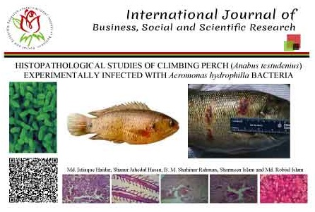
HISTOPATHOLOGICAL STUDIES OF CLIMBING PERCH (Anabus testudenius) EXPERIMENTALLY INFECTED WITH Aeromonas hydrophilla BACTERIA
Research Area: Fisheries and Marin Science
Volume: 03
Issue: 03
Page No: 143-148
Emailed: 7
Total Downloads: 1818
Country:

PDF View:
A study was conducted to assess the histopathology of climbing perch (Anabus testudenius) experimentally infected with Aeromonas hydrophilla bacteria. Anabas testudineus (koi) of 18g body weight were experimentally infected with Aeromonas hydrophila. Tissue samples from infected skin, muscle, gill, liver, spleen and kidney, in case of small fish whole diseased fingerlings will be preserved in 10% buffered formalin solution. Normal hepatocytes with no pathological infestation. Liver synocytes are of normal appearances. No bacterial colonies were observed. Liver of A. testudineus experimentally infected with A. hydrophila by OI method showing the haemorrhages (H) in the hepatic ducts. H & E × 40. Cross section of normal hematopoietic tissue with no abnormal condition of the renal tubules. Blood vessels were normal in appearance. Gill of normal A. testudineus where lamellae are unaffected. Both primary gill lamellae and secondary gill lamellae are arranged normal fashion.The pathological signs by experimental infections cioncided with those by natural infections. So, the bacteria A. hydrophila was suggested to be a pathogen of koi cultured in Bangladesh.